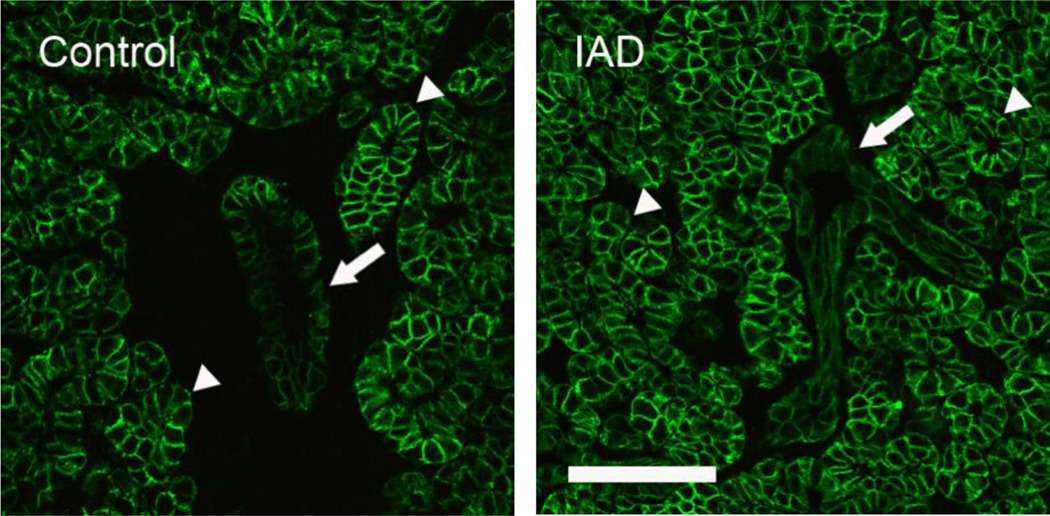
Fig. 5.
Immunofluorescence of NKCC1-IR. Control: NKCC1-IR was present at basolateral membranes of all acinar (arrowheads) and ductal cells (arrow), but the intensity was lower in ductal cells. IAD: NKCC1-IR in the acinar and ductal cells from IAD rabbits exhibited similar distribution pattern to that of control animals, with the staining intensity in ducts (arrow) was also much lower. Scale bar=50 µm.

