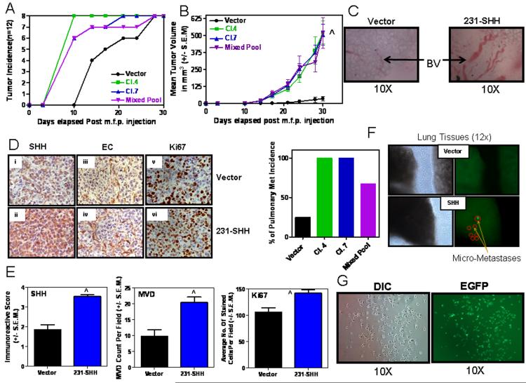
Figure 2. Constitutive SHH expression in MDA-MB-231 cells potentiates tumor growth and spontaneous metastasis.
200,000 cells were injected into the mammary fat pad of athymic nude mice. A) All mice injected with SHH-expressing breast cancer cell lines developed palpable tumors 10 days after injection. However, mice in the cohort of the vector control cells lagged behind. B) SHH expressing mice showed significantly increased tumor growth (^p < 0.0001 for all SHH-expressing groups relative to vector) and, C) prominently increased blood vessel (BV) formation in the primary tumors. D) The xenografts obtained from the MDA-MB-231-SHH expressing cells show a greater staining intensity for SHH (Panels i and ii), endothelial cells staining for lectin (Panels iii and iv) and Ki67 (Panels v and vi). E) Concomitant with staining intensity, the xenografts obtained from MDA-MB-231-SHH cells show greater immunoreactive scores for SHH (^p < 0.0001), microvessel density, MVD (^p = 0.0019) and Ki67 (^p = 0.0062). F) Tumors from SHH expressing cells also yielded a high incidence of pulmonary metastasis. Shown are representative photomicrographs of the lung images using the Stereoscopic Zoom Microscope (Nikon SMZ1500, Nikon Instruments Inc. Melville, NY) denoting visible light (left panels) and u.v. fluorescence (right panels) imaging that allows visualization of EGFP-expressing metastatic cells. G) The EGFP-expressing metastatic cells from the lungs were isolated in culture. Shown are representative photomicrographs in DIC and u.v. fluorescence.

