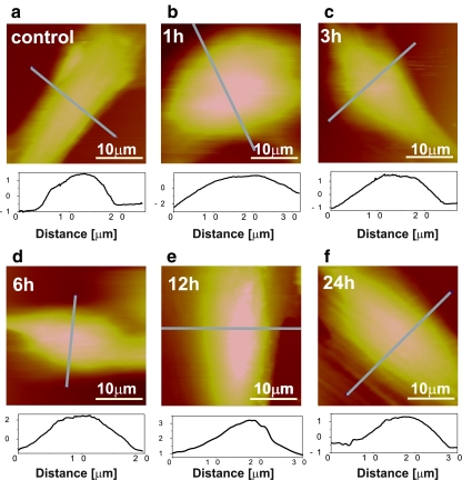
Fig. 5.
AFM images presenting 30×30 μm morphology scans of the HMEC incubated with TNF-α. The spindle-shaped control cell (a) was not exposed to cytokine. b–f Representative cells stimulated with TNF-α for 1, 3, 6, 12, and 24 h, respectively. Images revealed the change of cell shape: from spherical (1 h) to longitudinal for longer incubation periods

