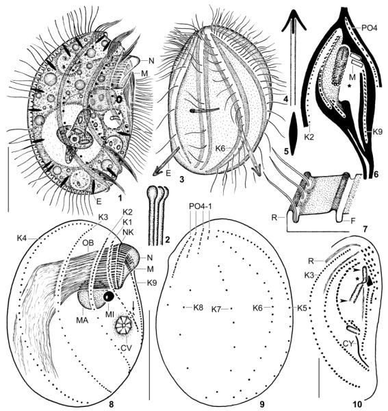
Fig. 1–10.
Leptopharynx bromelicola n. sp., macrostome specimens from life (1, 3–5), redrawn from scanning electron microscopy micrographs (2, 6, 7), after protargol impregnation (8, 9), and after Chatton–Lwoff silver nitrate impregnation (10). 1, 3. Right and left side view of representative specimens, the left one has just captured an euglenid flagellate. Note the conspicuous ridges and furrows as well as the comparatively densely ciliated kinety 6, a main difference to L. costatus, which has only two cilia in this kinety. 2. Distal end of oral basket rods. 4, 5. Exploded and resting extrusome (~ 4 × 0.8 μm). 6. Ridge pattern (black) of ventral side. The asterisk marks a rather deep concavity left and underneath the oral basket. The stippled area is also deepened but less than that of the deep concavity. The oral basket is covered by membranous material in the anterior half and oblique to the body’s main axis. 7. Ridge and furrow pattern of the cortex. 8, 9. Right and left side view of macrostome hapantotype specimen, length 52 μm. The arrow marks the oral primordium. 10. Ventrolateral view showing the narrow opening of the oral basket (arrowheads) and the anterior elongation of the basket (nose, asterisk). AM, adoral membranelles; CV, contractile vacuole; CY, cytopyge; E, resting and exploded trichocysts; F, cortex furrow; K1–9, somatic kineties; MA, macrostome; MI, microstome; N, nose; NK, nasse kinetosomes; OB, oral basket; PO1–4, preoral kineties; R, cortical ridge; Scale bars 15 μm (Fig. 10) and 25 μm (Fig. 1, 2, 8, 9).

