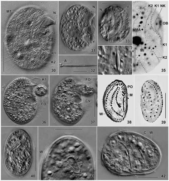
Fig. 30–42.
Leptopharynx bromelicola n. sp., trophic (30–37) and cystic (38–42) macrostome (30–37, 41, 42) and microstome (38–40) specimens from life (30–34, 36, 37, 40–42), after protargol impregnation (35), and Chatton–Lwoff silver nitrate impregnation (38, 39). 30, 31. Right side views of slightly squashed specimens, showing the typical nose produced by elongated oral basket rods. Note the numerous, sausage-shaped mitochondria (30, MT). 32, 34. Exploded and resting extrusome. 33. Left side view showing two deep furrows accompanied by ridges. 35. Right side view showing kineties 1 and 2 strongly diverging posteriorly. 36, 37. A specimen feeding on a resting cyst of a flagellate (Polytomella?). The cytoplasm is studded with lipid droplets. The cilia of kinety 1 (K1) form a membranous structure during feeding. 38–42. The resting cysts are covered by an about 1 μm thick wall, well recognizable in the squashed specimen shown in Fig. 42. The infraciliature, the oral basket, and some extrusomes are maintained, while the cilia are possibly resorbed. A, anchors; C, cortex; CV, contractile vacuole; CY, cytopyge; E, extrusomes; F, furrow; FO, food; K1, 2, somatic kineties; LD, lipid droplets; M, adoral membranelles; MA, macrostome; MI, microstome; MT, mitochondria; N, nose; NK, nasse kinetosomes; OB, oral basket; PO, preoral kineties; W, cyst wall. Scale bars = 5 μm (Fig. 32, 34), 15 μm (Fig. 35, 38–42), 25 μm (Fig. 36, 37), and 30 μm (Fig. 30, 31, 33).

