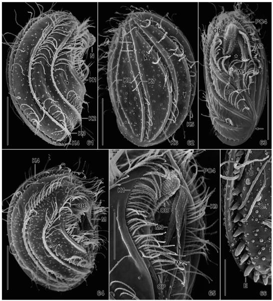
Fig. 61–66.
Leptopharynx bromelicola n. sp., macrostome specimens in the scanning electron microscopy. 61. Right side view, showing the typical nose, the ridges accompanying kineties 1–4, and the lack of cilia in mid-body. Kineties 1 and 2 diverge posteriorly, producing an elongate triangular area (asterisk). 62. Left side view, showing the kineties in deep furrows and accompanied by a ridge each right and left. The arrowheads mark circular accumulations of blebs possibly related to the extrusomes. 63, 65. Ventral and ventrolateral view, showing the oral ciliary pattern and the oral basket. The asterisk (63) marks the oral concavity. The arrow indicates the merging ridges of kineties 4 and 9. 64. Right side view of a broadly elliptical specimen showing the preoral kineties (arrowheads) occupying the ventral side, as in Leptopharynx costatus. 66. Right posterior corner, showing exploding extrusomes. C, cilia; E, extrusomes; K1–9, somatic kineties; M2, 3, adoral membranelles; N, nose; OB, oral basket; OP, oral primordium; PO4, preoral kinety 4. Scale bars = 5 μm (Fig. 66), 10 μm (Fig. 65), and 15 μm (Fig. 61–64).

