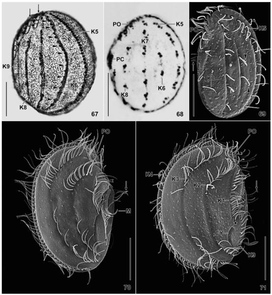
Fig. 67–71.
Leptopharynx bromelicola n. sp. (67) and Leptopharynx costatus (68–71) after Klein–Foissner silver nitrate impregnation (67, 68) and in the scanning electron microscopy (69–71). 67. Left side view, showing the dense cortical granulation and the comparatively dense ciliation of kinety 6 (arrowheads), a main feature of this species (bromelicola pattern). The arrows denote a narrow-meshed silverline pattern left of the preoral kineties and in the anterior portion of kinety 9. 68. Left side view of an Austrian population (from Foissner 1979), showing kinety 6 consisting of only two kinetids (costatus pattern). 69. Left side view of a German population, showing kinety 6 composed of two monokinetids. The arrow marks the cilia of the postoral complex. Numerals denote the preoral kineties. 70, 71. Right side views of a microstome and a macrostome specimen from a tank bromeliad of Mexico. The long axes of the oral baskets are marked by arrowheads. The arrows mark the ciliated portion of the postoral complex. CY, cytopyge; K1–9, somatic kineties; M, adoral membranelles; PC, postoral complex; PO, preoral kineties. Scale bars = 15 μm.

