Figure 2.

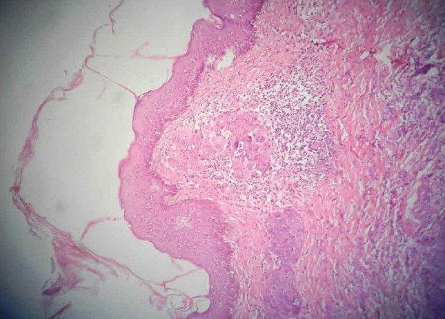
Histopathology; showing malignant cells forming glandular structures and solid sheets in the dermis (H and E stain, ×100)

Histopathology; showing malignant cells forming glandular structures and solid sheets in the dermis (H and E stain, ×100)