Fig. 1.
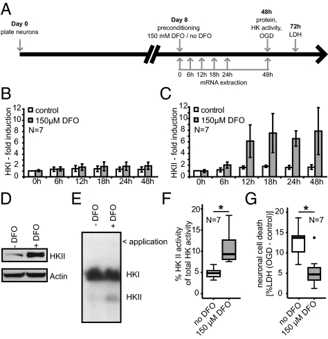
Hypoxia tolerance mediated by HIF-1–dependent activation of HKII in primary neurons. (A) Diagram of experimental paradigm. Analysis of HKI (B, E, and F) and HKII (C–F) expression in response to DFO treatment. (C) HKII expression was induced 12 h after hypoxia-mimicking treatment. After 48 h, (D) HKII protein and (E and F) enzyme activity were also increased. (B) mRNA expression and enzyme activity (E and F) of HKI remained unchanged. Representative immunoblot (D) and zymogram (E) 48 h after DFO treatment. (F) Summary of HKII activity using zymography after DFO treatment. *P = 0.007, unpaired two-tailed Student's t test. (G) DFO treatment reduced neuronal damage 24 h after OGD. *P = 0.006, unpaired two-tailed Student's t test. N indicates the number of independent experiments.

