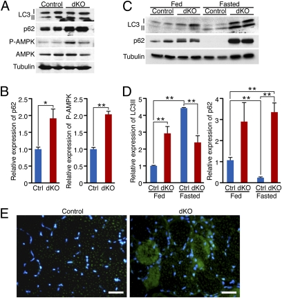
Fig. 2.
HDAC1 and HDAC2 regulate autophagy flux in skeletal muscle. (A) Western blot showing autophagy markers (LC3 I-II and p62), phosphorylated AMPK (P-AMPK), and total AMPK in skeletal muscle of neonatal control and dKO mice. Tubulin was used as loading control. (B) Densitometric analysis of Western blot showing p62 and P-AMPK expression in dKO relative to control (Ctrl) mice. Values were normalized to tubulin. n = 4. *P < 0.05; **P < 0.005. (C) Western blot showing LC3 I-II and p62 in skeletal muscle of 4-wk-old control and dKO mice, fed or fasted for 24 h. Tubulin was used as loading control. (D) Densitometric analysis of Western blot showing LC3 II and p62 expression in dKO relative to Ctrl mice. Values were normalized to tubulin. n = 6. **P < 0.005. (E) Immunostaining of histological sections of TA muscle for p62 (green) and nuclei (blue) at 7 wk of age. (Scale bars, 40 μm.)

