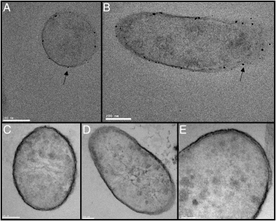
Fig. 1.
TEM images of thin sectioned T. potens. Intact cells were treated with Ag+ (A and B) or KCN before Ag+ (C–E) before thin-sectioning. (A) Ag precipitation (indicated by arrows) visible on the cell surface (A and B) is inhibited by the addition of KCN (C–E). A crystalline S-layer is not visible on the cell surface. Cells were stained with three stains: 0.5% Ruthenium red (polysaccharides), 2% Osmium tetroxide (lipids), and 2% Uranyl acetate (proteins and nucleic acids).

