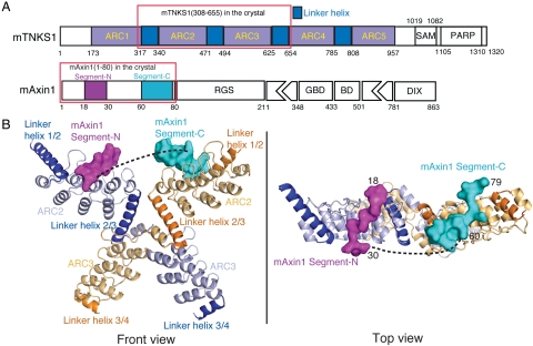
Fig. 1.
Overall structure of the TNKS(308-655)/Axin1(1-80) complex. (A) Schematic representation of the domain organization of mouse Tankyrase and Axin1. “ARC” stands for ankyrin-repeat cluster. Five ARCs are colored in gray. Linkers between them are colored dark blue. Domains SAM and PARP are also labeled. “GBD” stands for GSK3 binding domain; “BD” stands for β-catenin binding domain. RGS and DIX domains are also shown. Segment-N and –C of mAxin1(1-80) observed in the crystal structure are colored in magenta and cyan, respectively. (B) Overall structure of a complex between mTNKS ARC2-3 and mAxin1(1-80). Two copies of ARC2 and ARC3 in the dimer are shown in cartoon with light blue and light orange, respectively. Linker helices before and after ARC2 and ARC3 in two monomers are shown in blue and orange, respectively. The two Axin1 segments are shown in solid surface with magenta and cyan, respectively.

