Fig. 3.
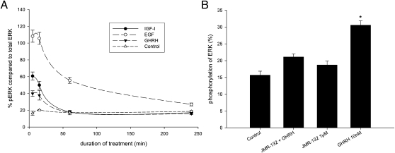
Activation of ERK in PC-3 human androgen-independent prostate cancer cells. (A) Time course of the phosphorylation of ERK related to total ERK protein in PC-3 prostate carcinoma cell line. Cells were treated with 10 nM IGF-I, EGF, and GHRH, which caused a significant increase in pERK after 5 min and 15 min compared with control (P < 0.001). EGF remained significantly elevated even after 4 h (P < 0.001). Statistical analysis was performed by Student's t test. (B) Phosphorylation of ERK in the PC-3 prostate cancer cell line related to total ERK protein. Stimulation of PC-3 cells with 10 nM GHRH caused a significant increase in p ERK (*P < 0.01) compared with control, whereas treatment with 1 μM of the GHRH antagonist JMR-132 showed no activation of ERK. Pretreatment for 30 min with 1 μM of JMR-132 almost completely abolished the activation of ERK by 10 nM of GHRH. Statistical analysis was performed by Student's t test.

