Fig. 2.
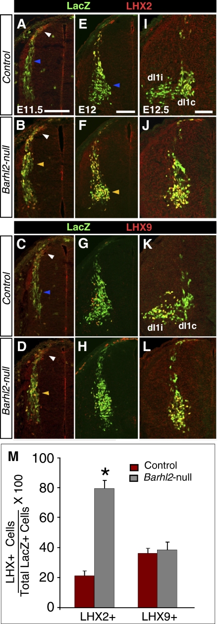
Ectopic LHX2 expression in dI1i neurons of Barhl2-nulls suggests dI1i-to-dI1c respecification. (A–D) In E11.5 Barhl2lacZ/+ controls, newly postmitotic neurons at the dorsal margin of the spinal cord (white arrowhead) express LHX2 and LHX9, but the ventrally migrating dI1i neurons express only LHX9 (blue arrowhead). In Barhl2lacZ/lacZ-nulls, dI1i neurons ectopically express LHX2 (yellow arrowhead). LHX2 at the dorsal margin and LHX9 are unchanged. (E–H) At E12.0, LHX2 continues to be ectopically expressed by dI1i neurons that have migrated to the medial deep dorsal horn (yellow arrowheads) in Barhl2-nulls. LHX2 expression in dI1c neurons and LHX9 is unperturbed. (I–L) At E12.5, Barhl2-expressing dI1 neurons segregate into lateral LHX2−/LHX9+ dI1i neurons and medial LHX2high/LHX9low dI1c neurons. In Barhl2-nulls, dI1 neurons fail to segregate into dI1i and dI1c groups. (M) Quantitation in I–L reveals an approximately fourfold increase (*P = 0.0001) in Barhl2-LacZ+ neurons expressing LHX2 in Barhl2-nulls. There is no change in Barhl2-LacZ+ neurons expressing LHX9 (P = 0.21). (Scale bars: 100 μm.)

