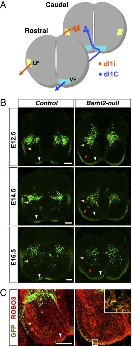
Fig. 3.
Genetic lineage tracing reveals a striking expansion of Barhl2+ axons in the VF. (A) Schematic. Medially located dI1c neurons project axons into the contralateral VF close to the midline. Laterally located dI1i neurons project axons into the ipsilateral LF. (B) Barhl2 genetic lineage tracing. GFP (green) immunohistochemistry on E12.5–E16.5 Barhl2cre/+; Z/EG controls reveals dI1i axons in the LF (yellow arrowheads) and dI1c axons in the VF (white arrowheads). In Barhl2cre/lacZ; Z/EG nulls, there is a drastic reduction of GFP+ fibers in the LF and a dramatic expansion of GFP+ fibers in the VF (red arrowheads). (C) GFP (green) and ROBO3 (red) double-immunolabeling indicates the contralateral origin of the ectopic GFP+ fibers spanning the mediolateral extent of the VF in Barhl2cre/lacZ; Z/EG nulls at E12.5. (Inset) Magnified view of a 0.4-μm-thick optical section of the boxed region. (Scale bars: 100 μm.)

