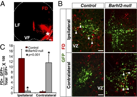
Fig. 4.
Dramatic increase in contralaterally projecting dI1 interneurons in Barhl2-nulls. (A) E13.5 spinal cord section depicting site of FD crystal placement (*) in the lateral VF and the extent of FD diffusion after 8 h of incubation during retrograde labeling. (B) Retrograde labeling from the lateral VF reveals FD+ back-labeled GFP+ neurons (white arrowheads) ipsilateral to the injection site in controls, as expected, and, strikingly, contralateral to the injection site in Barhl2-nulls. (C) Quantitation. There is a ∼20-fold decrease (P = 0.0001) and ∼26-fold increase (P < 0.0001) in GFP+ neurons back-labeled with FD ipsilateral and contralateral to the FD injection site in Barhl2-nulls, respectively. (Scale bars: 50 μm.)

