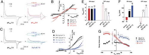
Fig. 5.
Cystine-dependent transient currents are carried by extracellular protons. (A and B) An individual oocyte was stepped to various potentials successively at pHout 7.5 and pHout 8.5. Cystine-dependent traces and charge-voltage relationships are shown in A and B, respectively. (C and D) Another oocyte was analyzed similarly in standard medium at pHout 7.5 and in deuterated medium at pDout 7.5. (E and F) Maximal charges (Qmax) and midpoint voltages (V0.5) for ON and OFF steps were determined (pHout 8.5, pHout 7.5) or extrapolated (pDout 7.5) by curve fitting. The extent of V0.5 shift observed between pHout 8.5 and 7.5 is consistent with the transfer of one proton from the extracellular medium to a fractional membrane depth of 0.64. (G) Decay times are plotted against the membrane potential to which oocytes were stepped. For the OFF steps, 3 steps from -130, +50 and +130 mV to -40 mV are plotted. Transient currents are slowed in deuterated medium, and in aqueous pHout 8.5 medium when Vm ≤ +40 mV. See also curve fitting analysis in SI Text. *P < 0.05 relative to pHout 7.5.

