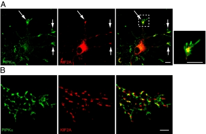
Fig. 2.
Endogenous localization of KIF2A and PIPKα. (A) Fluorescence images of cultured developing hippocampal neurons at 3 div double-stained with anti-KIF2A (red) and anti-PIPKα (green) antibodies. (Right) The boxed area is zoomed. KIF2A and PIPKα accumulated at the tips of neurites (arrows) and partially colocalized in growth cones. (Scale bars, 10 μm.) (B) Reconstructed signals for KIF2A and PIPKα in the growth cones detected by PALM. Fluorescence images of cultured developing hippocampal neurons at 3 div double-stained with anti-KIF2A (red) and anti-PIPKα (green) antibodies. (Scale bar, 1 μm.)

