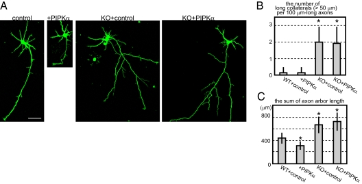
Fig. 5.
Overexpression of PIPKα in WT and Kif2a−/− neurons. WT or Kif2a−/− hippocampal neurons were transfected by GFP and either CFP (control) or CFP-PIPKα (+PIPKα) vectors at 2 d using a Ca2+-phosphate method, cultured for 1 d, and observed by GFP signals. (A) Representative images of each cell. (Scale bar, 50 μm.) (B) The number of collaterals longer than 50 μm was counted per 100 μm-long axons. Data are shown as mean ± SD; *P < 0.01, Student's t test; 13 neurons from four independent mice. (C) Total lengths of axons were calculated. Data are shown as mean ± SD; *P < 0.01, Student's t test; 12 neurons from four independent mice.

