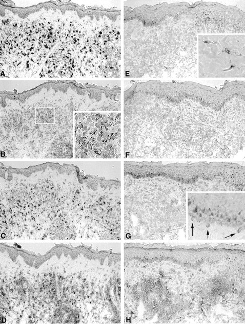
Figure 1.

Representative staining patterns with MAbs to PGL-I, LAM, 65 kd, and 36 kd in the lesional skin of untreated MB and PB patients. Immunoperoxidase staining of sequential sections of a MB lesion (BI 4+; A to D) and of a PB lesion (BI 0; E to H) with MAbs against PGL-I (A and E), LAM (B and F), 65 kd (C to G), and 36 kd (D to H). All sections were counterstained with hematoxylin. Immunoperoxidase single staining; magnification, ×125. Note the dark staining in the basal layer is due to melanin-containing melanocytes and not due to immunostaining.
