
Figure 3.
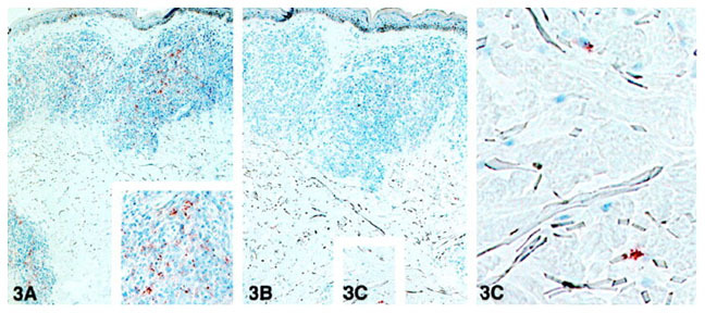
A and B: In situ detection of LAM and PGL-I in the course of the disease. Sequential sections of the lesion of a MB patient (1 year after the onset of the treatment, BI 2+) were stained with the MAb to LAM (A) and the MAb to PGL-I (B). C: Inset from B, showing scattered cells stained with the MAb to PGL-I. Immunoperoxidase single staining, hematoxylin counterstaining; magnification, ×40.