Figure 1.
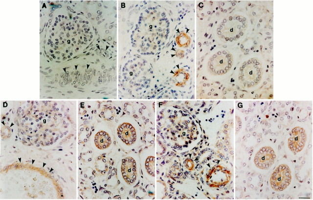
Localization of TGF-β1 and its receptors in normal developing kidneys. Antibodies were directed against TGF-β1 (A–C), TGF-βR1 (D and E), and TGF-βR2 (F and G) on sections of normal midgestation human kidneys. A: No immunoreactivity was detected when anti-TGF-β1 antibody was preincubated with excess peptide; vessels (arrowheads) and a glomerulus (g) are indicated. B and C: In sections in which the TGF-β1 antibody was not pre-absorbed, vessels were strongly immunoreactive, but fetal collecting ducts (d) did not show significant staining. D–G: In sections reacted with TGF-β1 receptor I and II antibodies, immunostaining was detected in fetal kidney vessels and collecting ducts. Scale bar, 15 μm.

