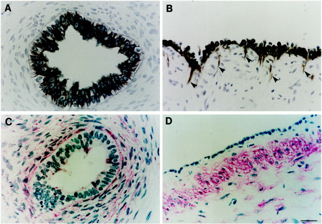
Figure 4.
Localization of cytokeratin and α-SMA in dysplastic kidneys. Dysplastic tubules are shown in A and C , whereas B and D show large cysts. Antibodies were directed against cytokeratin detected using diaminobenzidine (A and B), or α-SMA detected using fast red (C and D). No signal was detected on omission of primary antibody (not shown). A and B: All cells in dysplastic epithelia were positive for cytokeratin. A minor population of cytokeratin-positive cells were also detected in the interstitium proximate to large cysts (arrowheads in B); note the elongated appearance of these cells. C and D: α-SMA was detected in compact cells around dysplastic tubules and cysts. Rare cells were also weakly positive for α-SMA in dysplastic tubule epithelia in C. Scale bar, 15 μm.

