Figure 6.
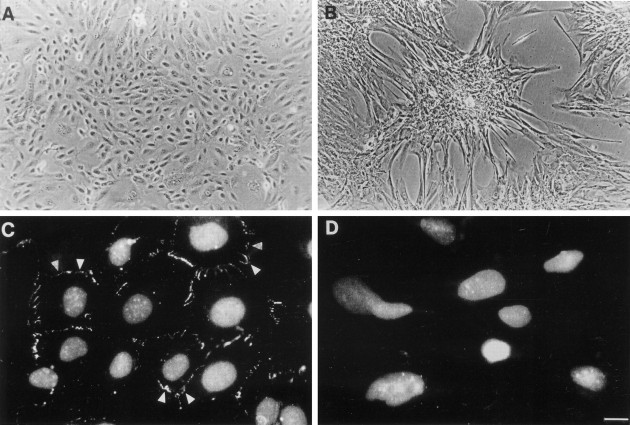
Effects of TGF-β1 on morphology and ZO1 expression. Gross morphology is shown in A and B, whereas C and D show ZO1 immunocytochemistry (with propidium-iodide nuclear counterstaining) of epithelial-like dysplastic cells cultured in either control medium (A and C) or with exogenous TGF-β1 (B and D). A: Cells cultured in control medium had an epithelial-like morphology in monolayer culture. B: Morphological changes were observed after exposure to 2.0 ng/ml TGF-β1 for 72 hours: multilayered aggregates formed in semiconfluent and confluent cultures and individual cells between aggregates became larger and developed filopodia and lamellipodia characteristic of a motile phenotype.34 C and D: In control medium ZO1 was immunolocalized to lateral cell junctions (arrowheads) but immunostaining at cell borders was lost after culture with TGF-β1. Scale bar: 40 μm (A and B); 15 μm (C and D).

