
Figure 2.
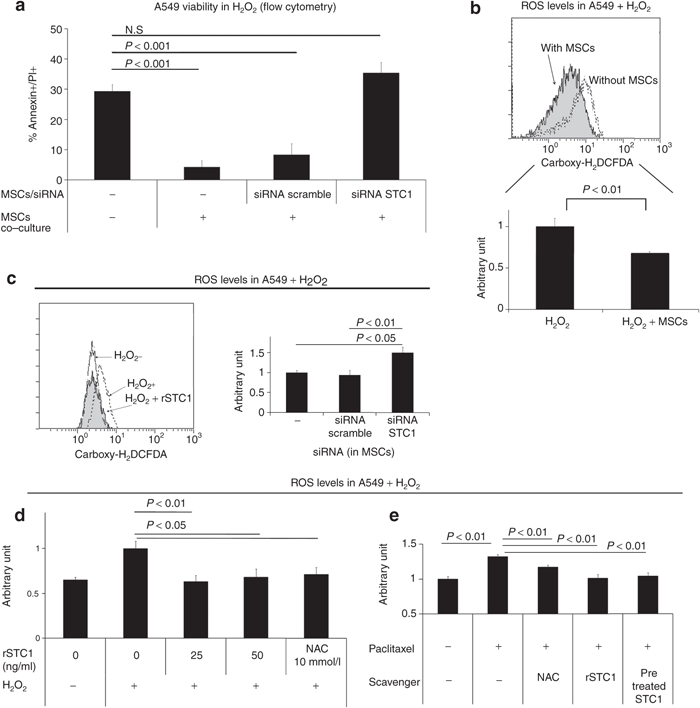
MSCs decreased ROS in H2O2-exposed A549 cells in a STC1-dependent manner. (a) A549 cells were incubated in culture medium containing H2O2 (100 µmol/l) with or without MSCs on a transwell filter. MSCs were transfected with a control or STC1 directed siRNA. Cells were assayed for viability using annexin V/PI staining. (b) A549 cells were incubated in a 6-well plate in culture medium containing H2O2 (100 µmol/l) with or without MSCs on a transwell filter. Four hours later, each culture was assayed for ROS using the dye, carboxy-H2DCFDA. Upper panel, representative flow cytometry data. Lower panel, quantification of ROS from three experiments. (c) MSCs transfected with scrambled or STC1-specific siRNA were co-cultured with A549 in medium containing H2O2 (100 µmol/l). Left panel: A549 cells were assayed for ROS using carboxy-H2DCFDA. Right panel: siRNA knockdown of STC1 in MSCs measured by real-time PCR. (d) rSTC1 was added to the A549 cell culture medium containing H2O2 (100 µmol/l). After 4 hours, A549 cells were assayed for ROS using carboxy-H2DCFDA. (e) NAC (N-acetylcysteine, 10 mmol/l), rSTC1 (50 ng/ml), and H2O2-pretreated rSTC1 (50 ng/ml) were added to A549 cells exposed to paclitaxel (5 µmol/l). Cells were assayed for ROS using carboxy-H2DCFDA. MSC, mesenchymal stromal cell; PI, propidium iodide; ROS, reactive oxygen species; siRNA, short interfering RNA; rSTC1, recombinant stanniocalcin-1.