Figure 6.

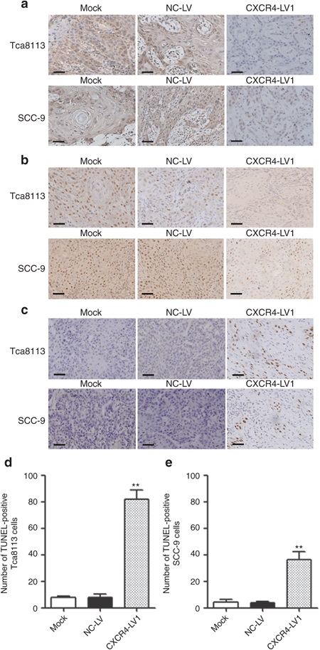
CXCR4 and Ki-67 IHC as well as TUNEL assay. (a) CXCR4-positive staining was located in the tumor nuclei and cytoplasm, and more positive cells could be seen in Mock and NC-LV cells than that in the CXCR4-LV1 Tca8113 and SCC-9 cells. (b) Ki-67-positive staining was located in the tumor nuclei, and more positive cells could be seen in the Mock and NC-LV cells than those in the CXCR4-LV1 Tca8113 and SCC-9 cells. (c) In the TUNEL assay, positive cells were located in the tumor nuclei. (d) The number of apoptotic cells significantly increased in CXCR4-LV1 Tca8113 cells compared with those in the Mock and NC-LV cells. (e) The number of apoptotic cells significantly increased in the CXCR4-LV1 SCC-9 cells compared with those in the Mock and NC-LV cells. Scale bar = 50 µm. Error bars indicate mean ± SD; **P < 0.01 IHC, immunohistochemistry.
