
Figure 6.
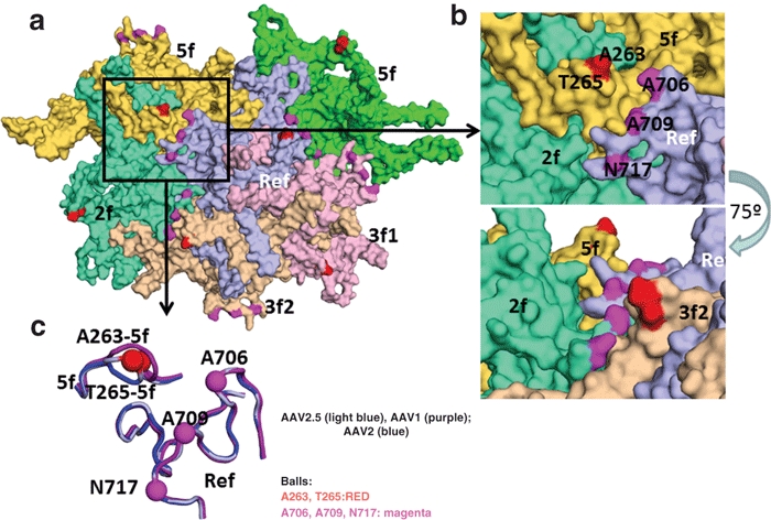
Topology of AAV2.5 virion. Surface topology view of the five VP monomers (in different colors) immediately surrounding the reference monomer in light blue. The symmetry operation that brings the monomer into contact is given in the labels. The positions of the AAV2.5 residue mutations are colored as in Figure 1b,c. (b) Top panel is a close up of the boxed region in the a. The bottom panel shows the same image rotated by ~75° and shows that the region containing the 263/265 amino acids are raised on the capsid surface. (c) Image showing a surface view (same as in Figure 1) of the loop containing the 263/265 region, VR III and VR IX (close to the 706, 709, 717 mutations) for AAV1 (purple), AAV2 (blue), and AAV2.5 (light blue). The positions of the AAV2.5 mutants are shown in the balls and labeled. AAV, adeno-associated virus.