
Figure 6.
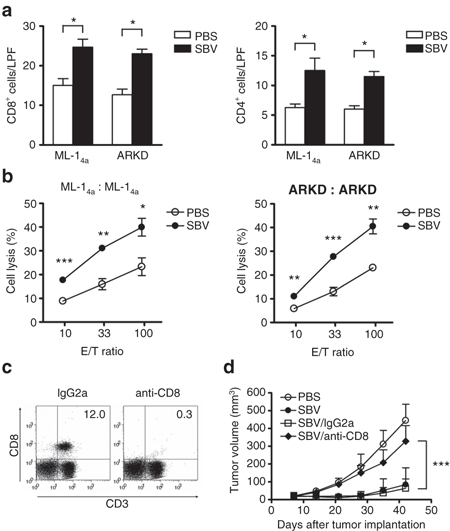
Sindbis virotherapy generates tumor-specific immune response. Tumor cells (5 × 10⁵/mouse) were subcutaneously inoculated in syngenic BALB/c mice on day 0. A single dose of SBV (1 × 10⁷ PFU/mouse) or PBS was intratumorally injected on day 7. Animals were killed on day 14 for the analysis of the Sindbis virotherapy-induced immune response. (a) Increased numbers of tumor-infiltrating CD8+ and CD4+ lymphocytes in both ML-14a and ARKD tumors treated with Sindbis virotherapy. Data presented are the quantification of IHC staining results from three low power fields (LPFs) per tumor and three tumors per group. (b) Enhancement of tumor-specific CTL response in the Sindbis virotherapy-treated animals. Mouse splenocytes were prepared from the mice bearing ML-14a or ARKD tumors. They were stimulated with mitomycin C-treated respective tumor cells for 5 days. The stimulated splenocytes were recovered and incubated with fresh ML-14a or ARKD cells, respectively, for 4 hours at the indicated E/T ratios. CTL activity was determined by lactate dehydrogenase release assays. (c) The efficiency of CD8+ T cells depletion. Animals were depleted of CD8+ T cells following a protocol as described in the Supplementary Materials and Methods by the anti-CD8 monoclonal antibody (53-6.72). An irrelevant IgG2a antibody was used as an isotype control. One day after the last antibody injection, the percentages of CD8+ T cells remained in the spleens were examined by flow cytometry. (d) The roles of CD8+ T cells in the Sindbis virotherapy-mediated antitumor activity. The growth of ARKD tumor was examined as described in the legend to Figure 5 in the SBV-treated animals which were depleted of CD8+ T cells or treated with an irrelevant IgG2a control antibody. *P < 0.05, **P < 0.01, ***P < 0.001, Student's t-test. CTL, cytotoxic T lymphocyte; PBS, phosphate-buffered saline; SBV, Sindbis virus.