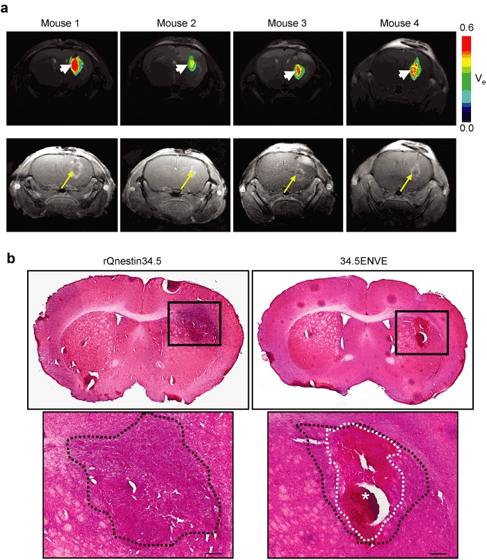
Figure 8.

Effect of 34.5ENVE on tumor necrosis. (a) Inverse spatial correlation of ve and contrast enhancing tumor area in 34.5ENVE-treated mice. Color-coded parametric images of ve in each of the four 34.5ENVE treated mice 3 days post-treatment (top) show increased ve (arrow head) in tumoral area that initially lacked contrast enhancement (arrow) immediately after Gd-DTPA administration in each of the four mice treated with 34.5ENVE. (b) Histologic analysis of necrosis in rQnestin34.5- and 34.5ENVE-treated brain tumors. In a parallel study, the above experiment was repeated and animals were sacrificed 3 days post-treatment for histological analysis of tumor-bearing brain. Representative hematoxylin and eosin (H&E) stained sections of rQnestin34.5- and 34.5ENVE-treated animals. Large necrotic area evident in 34.5ENVE-treated tumor (white dotted line and asterisks) is surrounded by viable tumor area (black dotted line). Bar = 200 µm.
