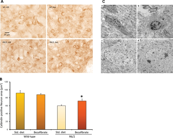
Figure 3.
Bezafibrate prevents neurodegeneration and increases mitochondrial density. (A) Calbindin staining in the striatum of 12-week-old R6/2 mice and their wild-type littermates on bezafibrate or standard diet. (B) Stereological analysis of calbindin-immunoreactive medium spiny neuronal perikarya in the striatum. The decrease in neuron size is significantly ameliorated by bezafibrate treatment. *P < 0.05 (n = 6 in each group). (C) Electron micrographs showing degenerated neurons in the striatum of R6/2 mice (a, b) and its amelioration by bezafibrate (c, d). a, b: Apoptotic neurons with condensed cytoplasm and abnormal nuclear shape showing margination and condensation of chromatin. The presence of large cytoplasmic vacuoles (bold arrow) and lysosome-like dense bodies is also noted. Degenerated mitochondria (light arrow) and lot of empty spaces can also be seen. c, d: In striata from bezafibrate-treated R6/2 mice, the cytoplasm of the neuron is preserved and the axonal and dendritic profiles in the neuropil are relatively intact. Scale bars, 2 μm. Magnification 10 000×.

