


Figure 2.
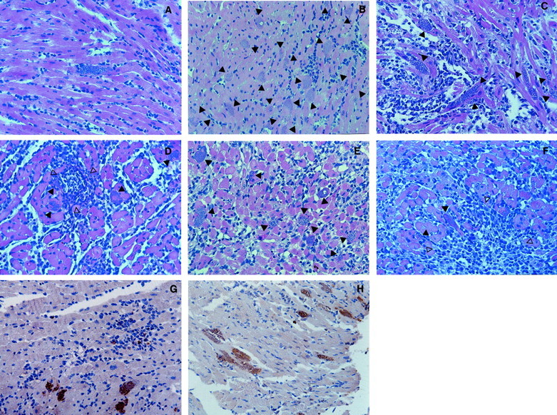
Illustration of T. cruzi parasitism and inflammation in cardiac tissue from WT and different KO mice infected with the Colombian strain of T. cruzi. A: One amastigote nest and mild inflammation is shown in one field of cardiac tissue from a C57BL/6 mouse at 15 days after infection (H&E; original magnification, 1 × 285). B: Twenty amastigote nests (closed arrow) and mild inflammation are shown in one field of cardiac tissue from an IFN-γ KO mouse at 15 days after infection (H&E; original magnification, 1 × 565). C: Seven amastigote nests (closed arrow) and intense diffuse inflammatory reaction are shown in one field of cardiac tissue from an iNOS KO mouse at 15 days after infection (H&E; original magnification, 1 × 285). D: Four amastigote nests (closed arrow) and intense diffuse as well as focal inflammatory reactions (open arrows) are shown in one field of cardiac tissue from a C57BL/6 mouse at 30 days after infection (H&E; original magnification, 1 × 285). E: Nine amastigote nests (closed arrow) and intense diffuse inflammatory reaction are shown in one field of cardiac tissue from a BALB/c mouse at 30 days after infection (H&E; original magnification, 1 × 285). F: Two amastigote nests (closed arrow) and intense diffuse as well as focal inflammatory (open arrows) reactions are shown in one field of cardiac tissue from an IL-4 KO mouse at 30 days after infection (H&E; original magnification, 1 × 285). Parasite-specific immunocytochemistry (brown staining) in cardiac tissue from IL-12 KO mouse at 50 days after infection (G) and IFN-γ KO mouse at 40 days after infection (H) with the Colombian strain of T. cruzi and treated with subcurative dose of benznidazole.