
Figure 2.
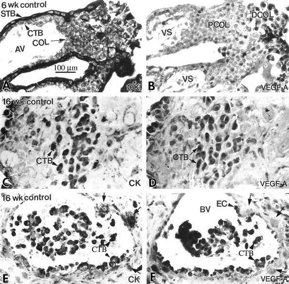
Cytotrophoblast staining for VEGF-A was up-regulated as the cells differentiated and invaded the uterus in situ. Serial paraffin sections of the maternal-fetal interface were stained with anti-cytokeratin (CK) to identify all of the trophoblast populations (A, C, and E), and with anti-VEGF-A (B, D, and F). Essentially the same staining pattern was observed during the first and second trimesters (ie, 6 and 16 weeks of gestation). A few cytotrophoblast (CTB) stem cells and cells in the proximal column (PCOL) region stained with anti-VEGF-A (B), but much more intense staining was observed in association with a majority of cytotrophoblasts in the distal regions of columns (DCOL) and with those that invaded the uterine wall (D). Cytotrophoblasts within the lumina of uterine blood vessels (BV) also exhibited intense staining (arrowheads), as did the maternal endothelial cells (EC; F). In contrast, some cytotrophoblasts in the vessel wall failed to react with anti-VEGF-A (arrows). The cells continued to stain for VEGF-A at term (see Figure 12B▶ ). AV, anchoring villus; STB, syncytiotrophoblast; VS, villous stroma; COL, cytotrophoblast column.