
Figure 3.
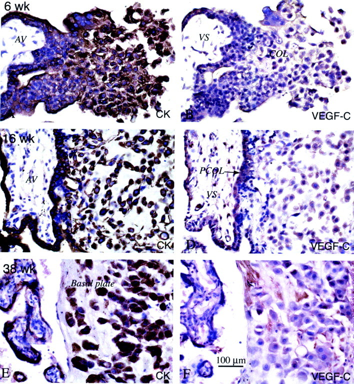
By the second trimester, cytotrophoblasts in all stages of differentiation stained with anti-VEGF-C in situ. Serial paraffin sections of the maternal-fetal interface were stained with anti-cytokeratin (CK) to identify the various trophoblast populations (A, C, and E), and with anti-VEGF-C (B, D, and F). In the first trimester (6 weeks), most of the cytotrophoblast populations stained with anti-VEGF-C, although cells in the distal portions of columns (COL) sometimes exhibited stronger antibody reactivity (B). In the second trimester (16 weeks), staining tended to be stronger than in the first trimester, with cytotrophoblast stem cells, cells in columns, and cytotrophoblasts within the uterine wall exhibiting similar levels of antibody reactivity (D). By term (38 weeks), little or no staining for VEGF-C was observed (F). AV, anchoring villus; VS, villus stroma; PCOL, proximal column.