
Figure 4.
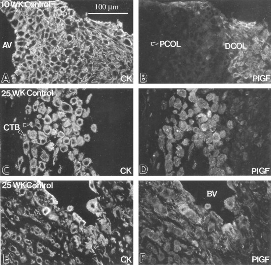
Cytotrophoblast staining for PlGF was up-regulated as the cells differentiated and invaded the uterus in situ. Frozen sections of the maternal-fetal interface in the first trimester (10 weeks) and the second trimester (25 weeks) were double-stained with anti-cytokeratin (CK; A, C, and E) and with anti-PlGF (B, D, and F). Essentially the same staining pattern was observed in the first and second trimesters. Staining for PlGF was first detected on cytotrophoblasts (CTBs) in the distal column (DCOL; B). Anti-PlGF reactivity was also observed in association with cytotrophoblasts that performed both interstitial (D) and endovascular invasion (F). At term cytotrophoblasts within the uterine wall either failed to stain or stained weakly with anti-PlGF (data not shown). AV, anchoring villus; PCOL, proximal column (arrowhead); BV, blood vessel.