
Figure 5.
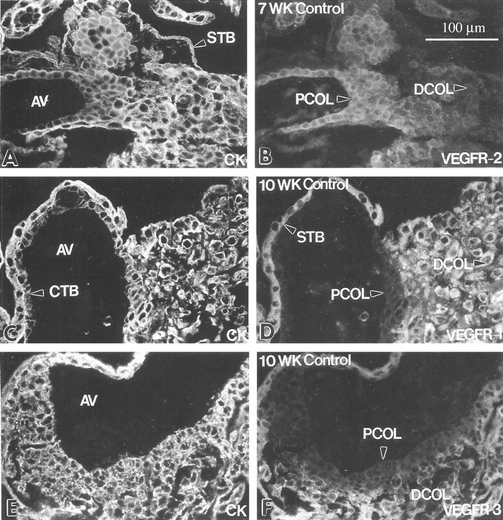
Cytotrophoblast subsets at the maternal-fetal interface differentially stain with antibodies that recognize individual VEGF receptors. Frozen sections of the first trimester maternal-fetal interface were double-stained with anti-cytokeratin (CK; A, C, and E) and antibodies that specifically reacted with one of three VEGF receptors (B, D, and F). Cytotrophoblast (CTB) stem cells and those in the proximal regions of cytotrophoblast columns (PCOL) stained with anti-VEGFR-2; little or no staining was detected in the distal column (DCOL) region and in association with cytotrophoblasts that invaded the uterine wall (B). Staining with anti-VEGFR-1 (D) and anti-VEGFR-3 (F) revealed a different pattern. Cytotrophoblast stem cells and those in the portion of the column immediately adjacent to the anchoring villus (AV) failed to react with either antibody, whereas cells in the rest of the column and within the uterine wall stained brightly with both. Syncytiotrophoblasts (STB) also stained for VEGFR-1 and VEGFR-3 (D and F). Essentially the same pattern was observed in the second trimester (data not shown). At term, only VEGF-R1 staining was detected in association with cytotrophoblasts (see Figure 13B▶ ).