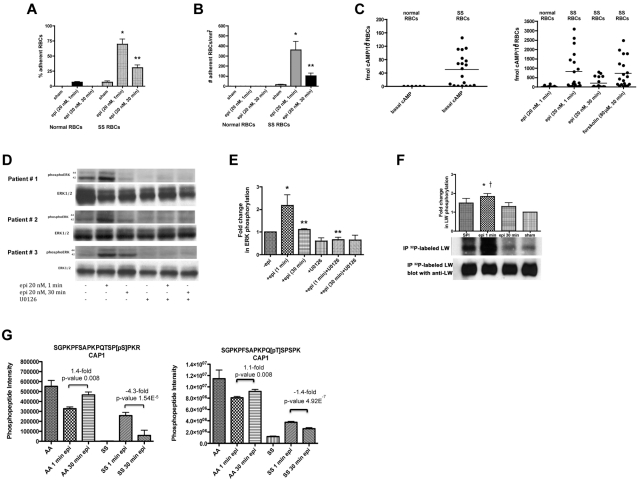
Figure 4.
SS RBC adhesion is associated with the extent of ERK activation. (A-B) Adhesion of SS RBCs to endothelial cells is related to the duration of cell stimulation with epinephrine. Adhesion of RBCs to HUVECs was tested in both intermittent flow and flowing condition assays, and results are presented as precent adherent RBCs at a shear stress of 2 dynes/cm2 and number of adherent RBCs/mm2, respectively. Normal and SS RBCs were sham-treated, or stimulated with epi for 1 minute or 30 minutes. *P < .001 compared with sham-treated SS RBCs; **P < .001 compared with epi-treated SS RBCs. Error bars show SEM of 4 different experiments. (C) cAMP production in SS RBCs is associated with the time of cell stimulation with epinehprine. RBCs were treated with IBMX (for basal cAMP levels), followed or not with epi (for 1 minute or 30 minutes) or forskolin. The specific effect of epi and forskolin on cAMP accumulation was obtained by subtracting basal cAMP levels from the total cAMP levels. The basal cAMP production and specific amounts of cAMP because of epi or forskolin stimulation were normalized as fmol cAMP/108 RBCs. (D-E) ERK phosphorylation is dependent on the time of SS RBC exposure to epinephrine. SS RBCs were sham-treated or treated with epi for 1 or 30 minutes, U0126, or U0126 followed by epi for 1 or 30 minutes. Immunoblots of RBC proteins with antibodies against ERK and phosphoERK (D) and quantitative analysis of the data presented as fold change in ERK phosphorylation (E) are shown. ERK underwent increased phosphorylation after 1 minute exposure to epi, whereas phosphorylation decreased with longer (30 minutes) cell exposure to epi (n = 4). *P < .01 compared with nontreated cells; **P < .01 and **P < .001 for epi-treated for 30 minutes and U0126+epi-treated for 1 minute versus cells treated with epi for 1 minute, respectively (E). (F) Inorganic 32P radiolabeled intact SS RBCs were incubated in the presence (lanes 1, 2, and 3) or absence (lane 4) of SPI, followed (lanes 2 and 3) or not (lane 1) by treatment with epi for 1 minute (lane 2) or 30 minutes (lane 3). The cpm are representative of 3 different experiments, calculated by subtraction of cpm present in a lane (not shown) containing immunoprecipitates using immunoglobulin P3 from cpm obtained using anti-LW (ICAM-4) mAb for immunoprecipitation under each set of conditions indicated. *P < .01 compared with sham-treated; †P < .05 compared with SPI + epi (30 minutes)–treated SS RBCs. (G) Prolonged cell exposure to epinephrine negatively affects phosphorylation of adenylate cyclase-associated protein 1. RBC ghosts isolated from SS and normal RBCs treated with epi for 1 and 30 minutes were enriched in phosphopeptides and then subjected to a label-free quantitative phosphoproteomics analysis. Phosphorylation of both serine and threonine on CAP1 in SS RBCs decreased with increased time (1 minute vs 30 minutes) of cell exposure to epi, whereas an increase in the abundance of these phosphopeptides was observed in normal (AA) RBCs after 30 minutes exposure to epi. Each data point is an average of 3 analytical replicate measurements with error bars indicating SD.

