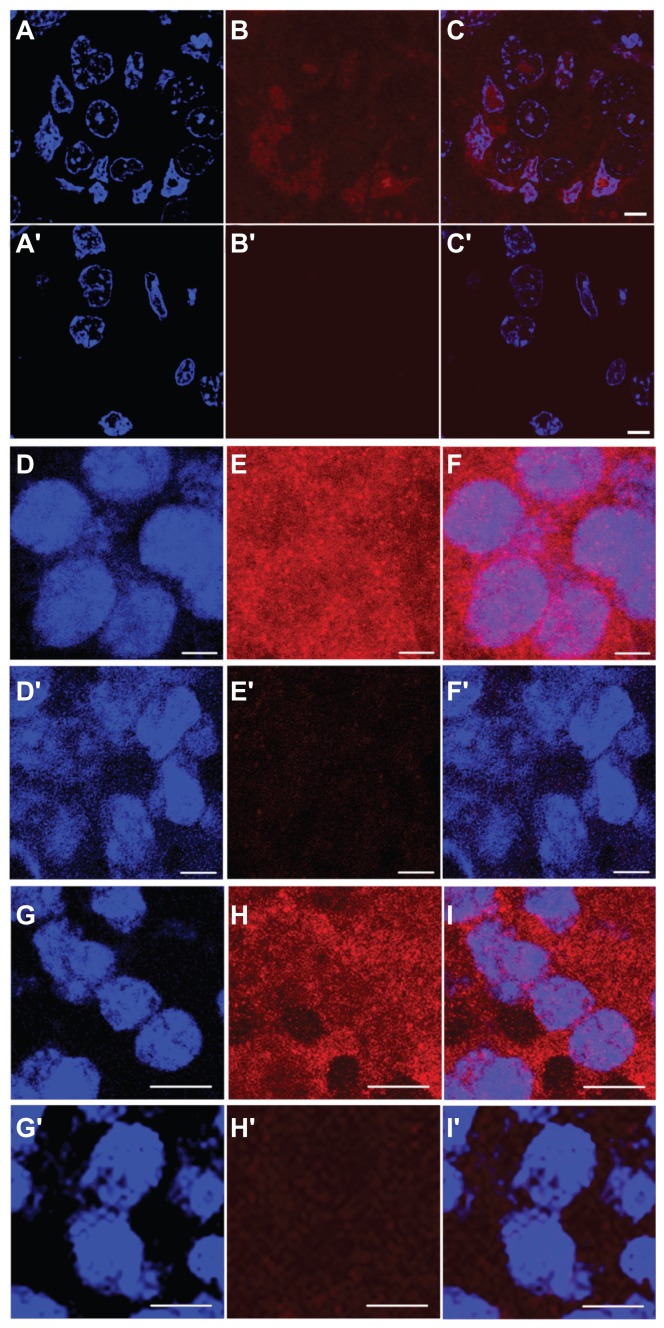
Figure 5.
Immunofluorescence analysis of in vivo subcellular localization of MNT 3 hours after intravenous injection in mice. (A–C) 2–3 μm tumor section (63× objective lens) from Balb/c ByJIco-nu/nu mouse bearing human A431 epidermoid carcinoma 3 hours after intravenous injection of chlorin e6-DTox-HMPNLS- EGF, (A′–C′) the same, but from the control mouse that was injected with saline. (D–F) 10 μm tumor section (63× objective lens) from C57 black/6J mice bearing murine B16-F1 melanoma injected with DTox-HMP-NLS-αMSH, (D′–F′) the same, but from the control mouse that was injected with saline. (G–I) 10 μm tumor section (63× objective lens) from DBA/2 mice bearing murine Cloudman melanoma S91 injected with DTox-HMP-NLS-αMSH, (G′–I′) the same, but from the control mouse that was injected with saline. (A), (A′), (D), (D′), (G), (G′) DAPI staining of cell nuclei (blue); (B), (B′), (E), (E′), (H), (H′) Alexa Fluor 555 staining for MNT (red); (C), (C′), (F), (F′), (I), (I′) overlay of cell nuclei fluorescence (blue) and MNT fluorescence (red). Scale bar 5 μm for (A–F) and for (A′–F′); scale bar 10 μm for (G–I) and for (G′–I′).
Abbreviations: MNT, modular nanotransporter; DTox, translocation domain of diphtheria toxin; HMP, the Escherichia coli hemoglobin-like protein; NLS, nuclear localization sequence; EGF, epidermal growth factor; αMSH, α-melanocyte stimulating hormone; SEM, standard error of mean; DAPI, 4′,6-diamidino-2-phenylindole.

