Figure 7.
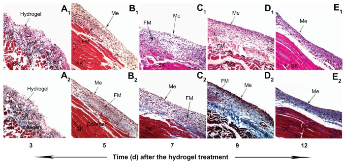
The fibrosis and the remesothelialization of the peritoneal wounds treated with PECE hydrogel. Three days after the treatment, a coating of the hydrogel on the injured surface of the abdominal wall, with infiltration of inflammatory cells (A1, 200×) and scattered collagen beneath (A2, 200×). Five days after the treatment, the layer of inflammatory cells composed mainly of foamy macrophages appeared, above which was a layer of spindle-shaped mesothelial cells (B1, 200×), and under which was a collagen layer (B2, 200×). Along with days, collagen- and fibroblast-rich tissues gradually increased and replaced the structure of the inflammatory cell layer (C1, C2, D1, D2, E1, E2, 200×).
Abbreviations: Me, mesothelial cell layer; SK, skeletal muscle; FM, foamy macrophages; PECE, poly (ethylene glycol)-poly (ɛ-caprolactone)-poly (ethylene glycol) (PEG-PCL-PEG).

