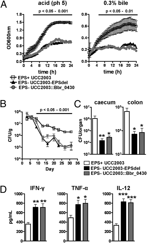
Fig. 2.
B. breve surface EPS protects against acid and bile, facilitates in vivo persistence, and modulates cytokine expression from stimulated splenocytes. (A) Growth curve of UCC2003 (EPS+), UCC2003-EPSdel (EPS−), and UCC2003::Bbr_0430 (EPS−) in de Man-Rogosa-Sharpe (MRS) pH5.0 or in MRS supplemented with 0.3% bile over 24 h at 37 °C. Data represent mean ± SD (B) BALB/c mice were treated orally with ∼1 × 10⁹ UCC2003, UCC2003-EPSdel, or UCC2003::Bbr_0430 on 3 consecutive days and bacterial numbers (CFU) in feces determined (data represent log10 CFU/g feces ± SD). (C) Organs were also removed on day 31 to determine CFU; columns show log10 CFU/organ (± SD). (D) Spleens were harvested from naïve BALB/c mice and stimulated with wild-type (EPS+), deletion, and insertion mutants (EPS−) B. breve strains at 1:1 ratio for ∼20 h. Cells were also stimulated with ConA (dotted line). Columns represent the mean ± SD stimulation indices of splenocytes from 10 mice from two independent experiments. Significance was determined relative to mice treated with UCC2003 (EPS+) at the same time point using the Kruskal–Wallis test followed by Dunn's multiple comparison test; *P < 0.05; **P < 0.01; ***P < 0.001.

