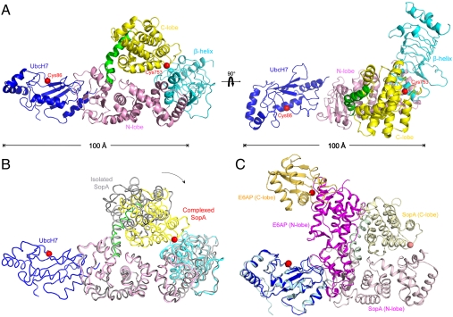
Fig. 1.
Crystal structure of the UbcH7/SopA complex. (A) Ribbon representation of the UbcH7 (blue) in complex with SopA (β-helix domain, cyan; N-lobe, pink; C-lobe, yellow; hinge helix, green). The two views differ by a 90° rotation. (B) Superposition of the isolated SopA (gray, PDB code: 2QYU) and SopA in the complex via the N-lobes (PDB code: 3SY2). An approximately 37° rotation of the C-lobes was observed about the hinge helix. (C) Comparing UbcH7/SopA and UbcH7/E6AP by superposition of the E2 molecules (blue and cyan). SopA is rendered in pink (N-lobe), yellow (C-lobe), and green (hinge helix). E6AP is shown in magenta (N-lobe) and orange (C-lobe). The catalytic cysteine residues are represented as red spheres.

