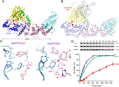
Fig. 3.
Crystal structure of UbcH7/NleL complex. (A) Ribbon representation of the UbcH7/NleL complex, same color theme as in Fig. 1. Catalytic cysteine residues are represented as red spheres. (B) Superposition of the isolated NleL (gray, PDB code: 3NB2 chain C) and NleL in the complex (colored as in panel A). An approximately 165° rotation of the C-lobes was observed about the hinge helix, and the catalytic cysteine residue C753 (gray and red spheres) is displaced by 45 Å. (C) F63 of UbcH7 interacts with different residues in SopA (left) and in NleL (right). (D) The ligase activities of wt and mutant NleL in multiturnover experiments, quantified by the percentages of mono-Ub usage. The error bars show the standard deviation from three independent experiments. Gel insets of the mono-Ub bands are also shown.

