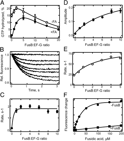
Fig. 4.
Effect of FusB on the dissociation of S. aureus EF-G from the ribosome. (A) GTP hydrolysis at conditions of EF-G turnover. Ribosomes (0.5 μM) and EF-G (0.5 μM) were reacted with [γ-32P]GTP (1 mM) without FA (○) or in the presence of FA (50 μM; ●) for 5 min at 25 °C. The extent of GTP hydrolysis was determined by TLC (30). (B) FusB-induced dissociation of ribosome⋅EF-G⋅mant-GDP⋅FA complexes. Ribosome⋅EF-G⋅mant-GDP complexes were formed in the presence of FA (200 μM) and rapidly mixed with FusB at increasing concentrations (0–5 μM, from top to bottom) at 37 °C. Complex dissociation was monitored by mant fluorescence. (C) Rates from B. Stopped-flow traces were evaluated by single-exponential fitting. (D) Amplitudes from B. (E) FusB-induced dissociation of ribosome⋅EF-G⋅BODIPY FL-GDP complexes. Complex dissociation was followed at 37 °C monitoring BODIPY fluorescence. Rates were determined by single-exponential fitting of stopped-flow traces. (F) Effect of FusB on ribosome⋅EF-G⋅BODIPY FL-GDP⋅FA complex formation. Complex formation was examined without FusB (●) or in the presence of FusB (2.5 μM; ■) at 37 °C, monitoring BODIPY fluorescence. For experimental details, see Materials and Methods.

