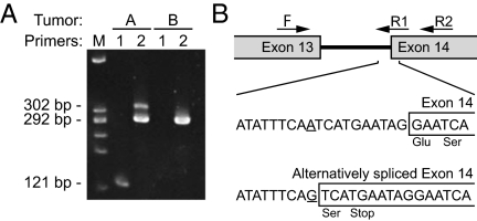
Fig. 2.
RT-PCR confirmation of alternative splice forms of the Apc transcript generated by a splice acceptor mutation in intron 13. (A) RT-PCR of tumors A and B using primers spanning intron 13. The PCR product from the normal splice form (primer set 2) is shown in tumor B (292 bp); it is also present in tumor A. Primer set 1 is specific to the alternative splice form created by the A–G transition mutation in intron 13. The 121-bp band is present only in tumor A. The differences in band intensity for primer set 2 in tumor A indicate that the alternative splice form is less abundant than the normal splice form. (B) Map and PCR primers spanning intron 13. Sequencing of the alternative splice form identified the sequence and position of the mutation that creates a splice acceptor.

