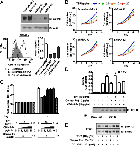
Fig. 4.
TSP1 inhibition of endothelial cell growth is reduced by CD148 knockdown or CD148-Fc. (A) HRMEC were plated in a six-well plate at a density of 50%. Lentivirus (1 × 10⁶ infectious units) encoding CD148-targeting or scramble shRNA was added to the cells with 5 μg/mL Polybrene (Santa Cruz Biotechnology). (Upper) Cells were harvested 72 h after infection, and CD148 expression was assessed by immunoblot analysis. Equal loading was confirmed by reprobing for β-actin. (Lower left) FACS analysis of CD148 expression in HRMEC treated with CD148-targeting (shRNA #1) or scramble shRNA. (Lower right) Fold change of the CD148+ cell fraction, compared with untreated HRMEC. (B) HRMEC treated with CD148-targeting or scramble shRNA lentivirus were plated in 98-well plates (2 × 10³ cells per well) 24 h after infection. Cells were starved for 12 h, and then TSP1 (0, 2, 10, 20 μg/mL) was added to the growth medium containing basic FGF (20 ng/mL). The medium was replaced at day 2 with fresh reagents. Cell number was assessed at the indicated time points. Data are means ± SEM of quadruplicate determinations. (C) TSP1 (10 μg/mL) was added to HRMEC with or without CD148-Fc (0, 1, 5, 10 μg/mL) or control Fc (1.2 μg/mL). Cell proliferation was assessed as in B. (D) HRMEC were plated in 100-mm dishes at a density of 30%, serum reduced [2.5% (vol/vol) FBS], and exposed to TSP1 (10 μg/mL) for 15 min with or without CD148-Fc (10 μg/mL) or control Fc (1.2 μg/mL). CD148 was immunoprecipitated using anti-CD148 or control IgG, and the washed immunocomplexes were assayed for PTP activity with or without 1 mM sodium orthovanadate (VO4). The amount of CD148 in the immunocomplexes was assessed by immunoblot analysis. The data show means ± SEM of quadruplicate determinations. *P < 0.01 vs. vehicle-treated cells. (E) HRMEC were treated as in D, and phosphorylation level of Erk1/2 was assessed by immunoblot analysis.

