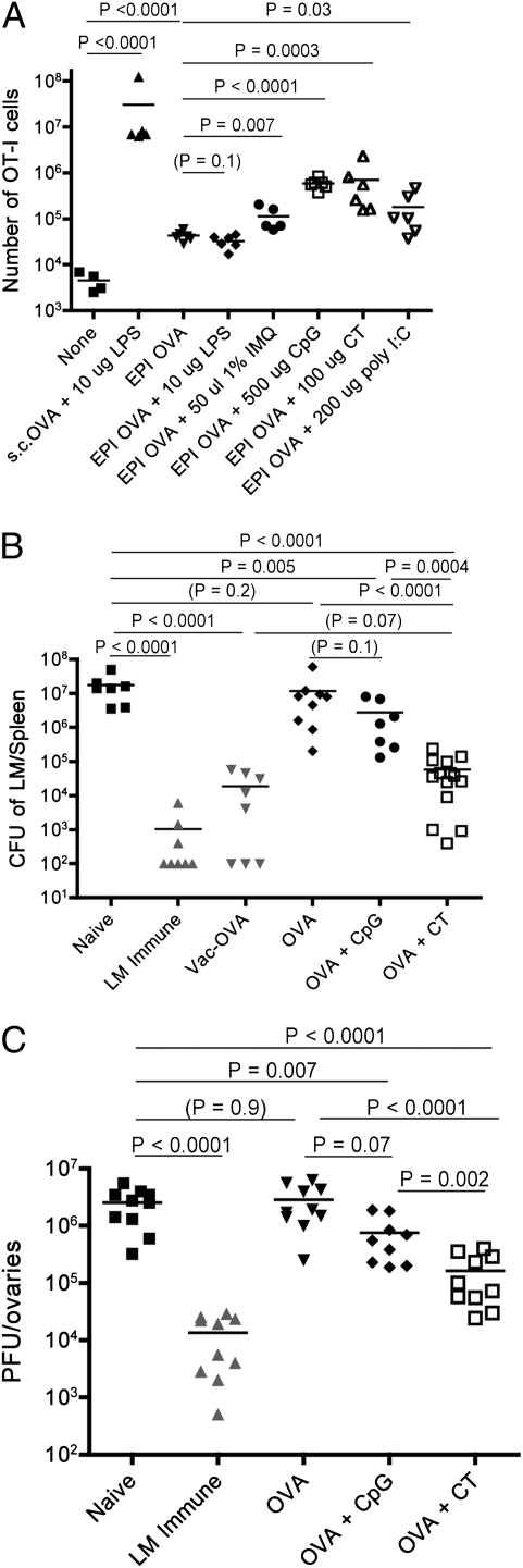
Fig. 1.
CT is a potent adjuvant for epicutaneous immunization of protective CD8 T cells. (A) C57BL/6 mice received 2.5 × 10⁵ CD44low OT-I cells 1 d before epicutaneous immunization (EPI), which involved administration of OVA protein with the indicated adjuvants on the ear skin. As a positive control, mice were primed via s.c. priming with OVA and LPS. Six days postimmunization, expansion of OT-I cells was determined by flow cytometric analysis of spleen and lymph nodes. Mice listed as “none” received OT-I cells but no immunization. The dose of each adjuvant is indicated. Each symbol represents an individual mouse. The data are compiled from two experiments and are representative of at least three similar experiments. (B and C) B6 mice were primed via epicutaneous vaccination as in A, except that OT-I cells were not transferred. Thirty days later the animals were boosted with the same immunization approach, and after an additional 30 d were challenged. (B) Mice were challenged with LM-OVA and protective immunity was assayed 5 d later. The data show LM-OVA cfu in the spleen of the indicated animals. As positive controls for protective immunity, mice primed with LM-OVA (“LM immune”) or VV-OVA (“Vac-OVA”) were also challenged. Data are compiled from three to four independent experiments. (C) Mice were challenged with VV-OVA and viral control was measured in the ovaries 3 d later. As a positive control, some mice were infected with LM-OVA at least 1 mo before VV-OVA infection (LM immune). These data are compiled from three individual experiments.

