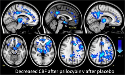
Fig. 2.
Decreased CBF after psilocybin (ASL perfusion fMRI). Regions where there was significantly decreased CBF after psilocybin versus after placebo are shown in blue (z: 2.3–3.7). Mixed effects analysis, z > 2.3, P < 0.05 whole-brain cluster-corrected, n = 15. LH, left hemisphere; RH, right hemisphere. Note, we observed no increases in CBF in any region.

