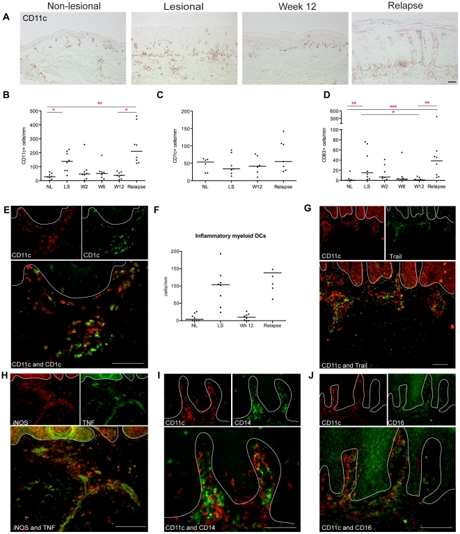
Figure 4. Increased inflammatory myeloid DCs in relapsed lesions.
(A) Representative immunohistochemistry and (B) counts of CD11c+ cells per mm in non-lesional skin (NL), lesional skin (LS), and in the index lesional plaque at weeks 2, 6 and 12 and time of disease relapse. (C) The numbers of CD1c+ cells did not change with treatment or relapse. (D) CD83+ mature DCs followed the same pattern as CD11c+ DCs. (E) There were many inflammatory myeloid DCs in the relapsed lesions, shown by immunofluorescence as CD11c+ cells (red) that were not co-expressing CD1c (and thus were not yellow), and (F) quantified as CD11c+ minus CD1c+ cells. (G) Inflammatory CD11c+ DCs were co-expressing TNFSF10/TRAIL. (H) TNF- and iNOS-producing DCs (TIP-DCs) were found in relapsed lesions. CD11c+ cells (red) co-expressing (I) CD14 and (J) CD16 (both green). Cells that co-express the two markers in a similar location are yellow in color. A white line denotes the dermo-epidermal junction. Bar is 100 µm.

