
Fig. 6.
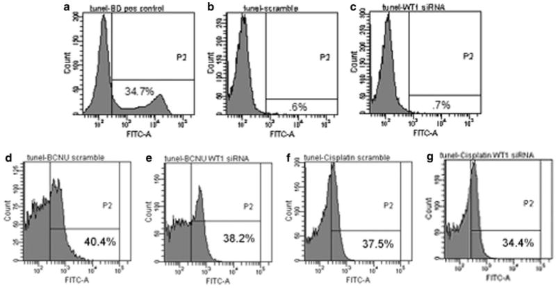
Representative flow cytometry experiments which indicate the amount of TUNEL staining in lymphoma and T98G cells. a The lymphoma cells were used as a positive control and show a classic second peak indicative of DNA fragmentation. Negative staining controls were performed without terminal deoxynucleotidyltransferase and used to set thresholds (<1%). Four days after transfection, T98G cells treated with (b) scrambled or (c) WT1 siRNA showed no significant increase in TUNEL staining. d–g Although treatment with BCNU and cisplatin alone resulted in DNA fragmentation, additional DNA fragmentation was not noted with WT1 downregulation. The percentage of cells with increased FITC-dUTP signaling is annotated in the lower right hand quadrant