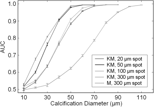
Figure 6.
As an example of the methods described in this work, the effect of the source spot size on calcification detectability was studied for the Konica-Minolta system geometry (denoted KM) and a typical mammography geometry (denoted M). These results show that, although the typical Konica-Minolta system configuration (source size of 100 μm) may offer somewhat better performance than mammography, improved detectability can be achieved when the source spot size is further reduced. Note that the mean glandular dose was held constant at 100 mRad for these studies and that imaging times varied.

