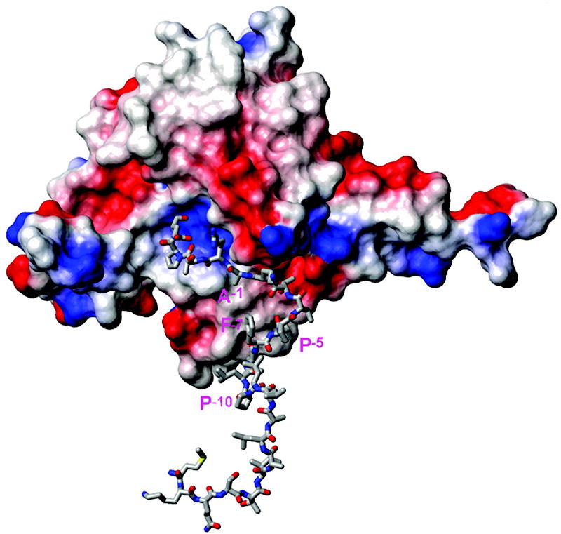
Figure 6.

Representative structure of the complex of SPase I Δ2-75 (PDB ID: 1KN9, shown in surface presentation colored by charge) with the KAP25 structure (represented by sticks) as obtained from HADDOCK docking. Residues Ala-1, Pro-5, Phe-7 and Pro-10 of KAP25 are marked in pink (using one letter code). Note that residues from the N-terminus to Pro-10 of the peptide are shown but do not interact with the enzyme and are likely to reside within the membrane.
